 Skip to content

Lipase: is an enzyme made up of proteins that play a critical role in the digestion and metabolism of lipids (fats). They break down dietary, endogenous and body fat.PMID: 30370868 PMID: 36302529 PMID: 30109644
There are different types of lipases that specialize in breaking down different types of lipids. For example:
Lingual (tongue) Lipase: Found in saliva secreted by the salivary glands, this enzyme begins the digestion of fats in the mouth and continues in the stomach by breaking down some triglycerides into smaller molecules called diglycerides.PMID: 2134569
Gastric lipase:The heat of the stomach liquidized the lipids. Both lingual lipase and gastric lipase continue breaking down fats, especially in the presence of an acidic environment.PMID: 30116546 PMID: 33710204
Digestion In Intestine
Bile salts:Bile salts are called biological detergents.PMID: 27031274
Short & medium chain fatty acids do not require bile salt for their absorption.
They absorbed directly in the intestinal cells, and they enter portal blood rather than lymph & transport to the liver bound to serum albumin.PMID: 10856722 PMID: 8785202
Bile salt is not absorbed along with digested product of lipids.PMID: 31689682
They reabsorbed in the lower part of the small intestine and retune to the liver.PMID: 35545671
Bile acids, can affect the pH at which pancreatic lipase activity is maximized.PMID: 1456492
Bile salt deficiency occurs in liver disease or due to obstruction in the bile duct.PMID: 33538845
Pancreatic Lipase – Digestion in Intestine:The Majority of fat digestion takes place in the small intestine, specifically in the duodenum, due to higher pH. Pancreatic Lipase enzyme is produced by the pancreas and is responsible for breaking them down into two fatty acids and a monoglyceride (dietary fats).PMID: 32344233 PMID: 27165048
This process takes place in the presence of bile acid, produced in the liver and stored in the gallbladder. Bile acid is released into the small intestine, which increases the surface area of fats breaking them into smaller droplets, making it easier for pancreatic lipase to work on them.PMID: 16431912 PMID: 29656109 PMID: 29080336
Cholesteryl esterase for breaking down Cholesteryl esters, Phospholipase for breaking down Phospholipids in the small intestine.PMID: 37161266 PMID: 22842588
Pancreatic enzyme deficiency occurs in pancreatitis or cystic fibrosis.PMID: 32761612 PMID: 36070542 PMID: 32168995
These lipid droplets are absorbed across the lining of the small intestine into the bloodstream. This absorption process is facilitated by specialized structures called villi and microvilli, which increase the surface area for nutrient absorption.
Fatty acids and monoglycerides are tacked together into triglycerides within the intestinal cells.
Triglycerides, along with other lipids and cholesterol, are packaged into structures called chylomicron.PMID: 21989069 PMID: 35296424
Chylomicron are too large (lipoprotein particles) to directly enter the tiny blood capillaries in the intestinal villi (small finger-like projections in the small intestine), where nutrient absorption primarily occurs. Due to their hydrophobic nature, they enter lacteals, specialized lymphatic vessels found in the villi to bypass the liver initially, avoiding potential congestion in the livers’ portal vein. Eventually merge with larger lymphatic ducts, the thoracic duct (one of the major lymphatic ducts), connects to the subclavian vein near the neck through which chylomicron eventually enters the bloodstream at a point closer to the heart. It is used for energy or stored by muscle and adipose (fat) tissue.PMID: 31361624 PMID: 30419164 PMID: 20961306 PMID: 26868089 PMID: 30020599 PMID: 29510243
Some leftovers (undergone partial breakdown) of chylomicron may reach the liver after they deliver triglycerides to peripheral tissues.PMID: 2536272
The liver can take up these remnants, and the lipids they contain, including cholesterol and fatty acids, can be processed into other lipoprotein for further transport or storage.PMID: 35331166 PMID: 21169224
Hepatic Lipase: Produced by the liver, this enzyme acts on triglycerides and phospholipids present in various lipoproteins, breaking them down into fatty acids and glycerol.
Chylomicron :(Greek chylo, means juice or milky fluid, and micron, means small particles)
Chylomicrons are the largest and least dense of the lipoproteins, and are synthesized in the intestines.
They carry dietary lipids, including triglycerides, phospholipids, cholesterol, and fat-soluble vitamins and transport from tissues to cell where they are either used for energy or stored in adipose tissue.PMID: 25974693
They contain only 1-2% protein, 85-88% triglycerides, ~8% phospholipids, ~3% cholesterol esters, ~1% cholesterol and apolipoprotein.
They are released into the lymphatic system and eventually enter the bloodstream. They are too large to pass through the capillary walls and are broken down into remnants after releasing their triglycerides.PMID: 31361624
After you consume a meal containing fat-soluble vitamins, such as vitamin A, D, E, or K, the vitamins are absorbed into the intestinal cells. Within these cells, the fat-soluble vitamins are packaged into chylomicron.
Vitamin A (Retinol): Chylomicron transports vitamin A from the intestines to the liver and other tissues.PMID: 12701068
Vitamin D: Chylomicron transports vitamin D from the dietary sources to the liver, where it’s converted into its active form.PMID: 30857638
The core of a lipoprotein particle is composed of hydrophobic lipids, mainly triglycerides and cholesterol esters. PMID: 21131533 PMID: 18261683
Surrounding the core is a surface monolayer which is hydrophilic (water-attracting) facing outward and a hydrophobic (water-repelling) facing inward, made up of a combination of phospholipids and free cholesterol molecules.
Within the surface of the monolayer there are attached proteins known as Apolipoprotein.PMID: 26095027
The key difference between them is chemical structure.
Molecular Structure
Fats in which the fatty acid chains have mostly single bonds between carbon atoms, allowing them to pack closely together, forming a solid structure and resulting in a Solid state at room temperature. Hence, they are also known as saturated fats, examples of fats include butter and Ghee.
In other words, Saturated fats have no double bonds between carbon atoms in their fatty acid chains. They are “saturated” with hydrogen atoms.
Saturated fats are solid at room temperature, shelf life is high, have high melting point, Rancidity is low and Unsaturated fats are usually liquid at room temperature, shelf life is low, have low melting point, Rancidity is high.PMID: 25979504
DNL primarily synthesized Saturated fats.
On the other hand, Fats in which the fatty acid chains have mostly contain one or more double bonds between carbon atoms, which prevent them from packing tightly together and resulting in a liquid state at room temperature. Hence, they are also known as unsaturated fats. Examples of oils include vegetable oil.
In other words, Unsaturated fats have two or more bonds in their fatty acid chains, which makes them twist and makes it hard for molecules to fit together tightly.PMID: 36537550
Unsaturated fats with specific double bond configurations, such as omega-3 and omega-6 fatty acids, must be obtained from the diet because the human body lacks the enzymes necessary to create these double bonds from scratch.PMID: 29715470
Unsaturated fats are produced through desaturation reactions, which introduce double bonds into the fatty acid chains. These reactions can occur in the body or, in some cases, be obtained directly from the diet.PMID: 11681705 PMID: 30764880
The distinction between single and double bonds lies in the number of electron pairs (Sharing of Electrons) shared between two atoms.
Single bonds allow atoms to share two electrons, one from each atom, resulting in a stable electron configuration.
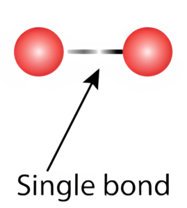
whereas In a double bond, two electrons are shared between the two bonded atoms. This sharing of electrons creates a strong attraction that holds the atoms together. These different types of bonds play a critical role in determining the reactivity of molecules in chemistry.


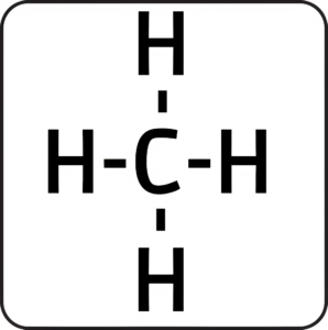
Basically, in a hydrocarbon chain, such as those found in fatty acids, carbon atoms can form four single bonds with other atoms. These bonds can be with hydrogen atoms or other carbon atoms.
In the case of Saturated, fats are composed of fatty acids with hydrocarbon chains in which every available carbon-carbon bond is a single bond.
Since carbon atoms can form up to four single bonds, in saturated fats, each carbon atom is bonded to as many hydrogen atoms as possible.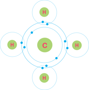
Whereas In the case of unsaturated fats, there is at least one double bond in the hydrocarbon chain of the fatty acid. Because a double bond forms, carbon atoms in the chain can’t bond with as many hydrogen atoms. Instead, they are bonded to fewer hydrogen atoms compared to carbon atoms in a saturated chain.
 Saturated fats are “saturated” with hydrogen atoms. In other words, every available carbon bond in the fatty acid chain is fully occupied by a hydrogen atom. The absence of double bonds between carbon atoms results in a straight, linear structure for the fatty acid chain, allowing the molecules to pack closely together. As a result, saturated fats are typically solid at room temperature.
 Whereas in unsaturated fat, the presence of double bonds reduces the number of hydrogen atoms attached to the carbon atoms, This prevents the molecules from packing as closely together. In other words, unsaturated fats are called unsaturated not because they completely lack hydrogen atoms, but because they have fewer hydrogen atoms in the locations where the double bonds are present.
Unsaturated fats are typically liquid at room temperature. The degree of fluidity depends on the number and placement of double bonds.
In short, unsaturated fats have double bonds in their hydrocarbon chains because of the sharing of two pairs of electrons between carbon atoms. This double bond creates a kink in the chain and results in different physical and health properties compared to saturated fats.
Why has Nature created saturated fats with a single bond and unsaturated fats with a double bond?
Why is saturated fat important?
Cells have lipid bilayer membranes that contain both saturated and unsaturated fats.PMID: 30472080
Saturated fats have straight, compact structures (fatty acids) in which all carbon-carbon bonds are single bonds (no double bonds). This absence of double bonds means that each carbon atom in the fatty acid chain is saturated with hydrogen atoms, hence the term “saturated fats.” PMID: 1608565
Because there are no double bonds to introduce twist or bends in the carbon chain, the carbon atoms in saturated fatty acids are linked together in a straight, linear fashion. This straight structure allows them to pack tightly together, making cell membranes more stable, rigid and less permeable. This stability is crucial for maintaining the integrity and rigidity of cell membranes.PMID: 29543154 PMID: 31329416
Omega-6 and omega-3 polyunsaturated fatty acids, contribute to different depths in bilayer membranes, affecting membrane processes. The balance between these two types of fatty acids in the diet has a significant influence on membrane composition and can alter essential physiological capacities of animals and humans. Hence, they play a crucial role in the function of biological membranes.PMID: 33914036 PMID: 27074681
DIAGRAM
Unsaturated fats, on the other hand, have twists in their structure due to the presence of double bonds between carbon atoms. These twists prevent them from packing closely together, making cell membranes more fluid, less stable and permeable. While some degree of fluidity is essential for membrane function, too much fluidity can compromise (bacterial growth and peptidoglycan synthesis) membrane stability.PMID: 36121000 PMID: 3060983 PMID: 32662773 PMID: 37277191
The synthesis of steroid hormones occurs within specialized cellular structures called the endoplasmic reticulum and mitochondria. The presence of cholesterol alone is not sufficient for hormone synthesis. Enzymes within the endoplasmic reticulum and mitochondria require a stable membrane environment to facilitate the complex chemical reactions involved in converting cholesterol into steroid hormones.
The endoplasmic reticulum and mitochondria structures have membranes composed of particularly phospholipids containing saturated fatty acids. These stable, saturated fat-rich membranes provide a suitable environment for the enzymes involved in steroid hormone synthesis to function optimally.PMID: 37207805 PMID: 2223867
The stability and rigidity provided by saturated fats in cellular membranes are essential for creating an optimal environment for the enzymes responsible for hormone synthesis. Unsaturated fats, due to their more fluid nature, do not provide the same structural stability required for these processes. Therefore, even though both saturated and unsaturated fats contain cholesterol, it is the structural properties of the surrounding lipid membranes that play a crucial role in facilitating steroid hormone synthesis.
Before that, we need to understand how cholesterol plays a role in stability and how cholesterol and saturated fat and unsaturated fat together play a role in stability.
Cholesterol molecules help regulate membrane fluidity by preventing the fatty acid chains from packing too closely together (in the case of saturated fats) or separating too far apart (in the case of unsaturated fats).
Mechanism
Cholesterol acts as a “buffer” in the membrane, preventing the fatty acid chains of the phospholipids from becoming too tightly packed at low temperatures or too loosely spaced at high temperatures, which helps maintain membrane fluidity and flexibility.
Saturated fats chains are relatively straight and can pack closely together in the lipid bilayer of a cell membrane by dominating the composition of phospholipids in the membrane, the membrane becomes more rigid and less fluid.
Cholesterol molecules inserted into the lipid bilayer among the saturated phospholipids act as “spacers.” They occupy space between the fatty acid chains, preventing them from packing too closely together. This increases the spacing between phospholipid molecules and, as a result, maintains membrane fluidity.
When unsaturated fats are predominant in the membrane, the presence of twists or bends in the fatty acid chains increases the space between phospholipid molecules, making them less orderly and promoting membrane fluidity.
Cholesterol prevents the fatty acid chains from separating too far apart, which helps restrict excessive membrane fluidity and effectively stabilizes the membrane.
In both cases, cholesterol acts as a “fluidity buffer.” It adjusts the membrane’s properties to maintain an optimal balance between rigidity and fluidity. This adaptability is essential for the proper functioning of cell membranes because it allows cells to function effectively under varying temperature conditions. Cholesterol ensures that the membrane remains functional and stable, preventing it from becoming too rigid in cold temperatures or too fluid in warm temperatures.
Overall, cholesterol’s role in regulating membrane fluidity is critical for maintaining the integrity and functionality of cell membranes, regardless of whether they are composed of saturated or unsaturated phospholipids.
Moreover, Cholesterol primarily influences membrane fluidity by regulating the arrangement of phospholipids, not fatty acids. Cholesterol molecules are placed among the phospholipids in the lipid bilayer of cell membranes.
The lateral structure of the bilayer is influenced by the properties of the lipid components, such as cholesterol and different phospholipids.
In saturated fats, they are broken down into individual fatty acids, including palmitic acid and stearic acid.
Primarily, palmitic fatty acids serve as substrates for the synthesis of VLDL in the liver; Hence, it has shown to increase the liver’s production of LDL cholesterol.
The body produces palmitic acid by releasing DNL in the liver, which in turn releases LDL cholesterol. Similarly, dietary palmitic acid from in Beef, pork, lamb, butter, cheese, whole milk, Palm Oil involved in Processed Foods and cooking oils. Cocoa butter from Chocolate, Coconut, macadamia (Nuts and seeds), shrimp and tilapia is involved in the production of LDL cholesterol. Combined, this process makes LDL cholesterol go up faster.
Additionally, saturated fats can reduce the liver’s ability to remove LDL cholesterol from the bloodstream, further contributing to elevated levels.
Elevated LDL cholesterol can lead to the down regulation of LDL receptors on cell surfaces. This means that cells become less efficient at taking up LDL particles from the bloodstream. As a result, LDL particles can circulate in the bloodstream for longer periods.
The body Produces (synthesizes) new fatty acids from non-lipid sources, such as carbohydrates (primarily glucose) and, to a lesser extent, amino acids. This process primarily occurs in the liver and, to some extent, in adipose (fat) tissue.
The process begins with the conversion of glucose (glycolysis) into Pyruvate, Pyruvate is then converted into acetyl-CoA (a central molecule in metabolism and can be derived from glucose and amino acids) through a series of enzymatic reactions.PMID: 18477307 PMID: 32152547 PMID: 32761568 PMID: 36892956 PMID: 26443368 PMID: 30409761 PMID: 17560727
Acetyl-CoA is converted to malonyl-CoA (the key building block for fatty acid synthesis) by enzymatic reaction. This step is considered the rate-limiting step in de novo Lipogenesis and is highly regulated. On the other hand, Malonyl-CoA also serves as an inhibitor of carnitine palmitoyltransferase I (CPT-1), an enzyme involved in fatty acid transport into mitochondria for oxidation. PMID: 36459649 PMID: 35810180 PMID: 31040353 PMID: 36917141
Malonyl-CoA goes into fatty acid production and by which saturated fat, also known as palmitic acid is produced, which then gets converted to triglyceride with the help of glycerol. Now from where does glycerol come. So Pyruvate also gets converted into glycerol and hence, in this way, with the help of saturated and glycerol, triglycerides are produced. This process is also known as esterification. The body can also get glycerol from other sources, such as dietary fat and adipose tissue. PMID: 4152155 PMID: 28827624
The newly synthesized triglycerides are then released in the bloodstream as a Lipoprotein.
While the body can synthesize palmitic acid, It can be obtained directly from the diet through the consumption of foods like meat, dairy products, and certain vegetable oils.PMID: 24731645 PMID: 25764297
De novo Lipogenesis is tightly regulated to prevent excessive fat production. Key regulatory factors include hormonal signals, such as insulin, which promotes DNL, and glucagon, which inhibits it. However, DNL can become more significant under certain conditions, such as high carbohydrate diets or when (calories) energy intake exceeds expenditure, leading to fat accumulation.PMID: 36976708 PMID: 36892956 PMID: 12499321 PMID: 12037719
DNL occurs in various tissues, including the liver and adipose tissue.PMID: 32201132
Acetate, leucine, and isoleucine promotes DNL.PMID 32201132.
Question raised here, BCAA is linked with Insulin Resistance, is it because of leucine and isoleucine promotes DNL? Click here to check.
It serves several essential functions in the human body and other organisms.
Energy Storage: Conversion of excess glucose and other non-lipid into fat and storing in adipose tissue.PMID: 25973385
Energy Regulation:In response to a high-carbohydrate meal, insulin promotes the conversion of glucose to fatty acids through DNL, which helps regulate blood glucose levels by removing excess glucose from the circulation.DNL plays a role in energy homeostasis.
Evolutionary Advantage: The ability to convert excess dietary carbohydrates into stored fat through DNL provided a survival advantage during times of food scarcity.PMID: 33730548
Lipoprotein Lipase:Lipoprotein lipase (LPL) is primarily responsible for the breakdown of triglycerides in circulating lipoproteins (LDL, VLDL & HDL), glycerol, and free fatty acids.
LPL has a dual mechanism of action, depending on its location and function in different tissues of the body.PMID: 29453968 PMID: 33277156
It is found at the highest levels in adipose tissue (fat cells) where it serves in the uptake of dietary fat and promotes fat storage during high calorie intake. On the other hand, during low calorie intake it helps to breakdown fats into glycerol, and free fatty acids.PMID: 2685199 PMID: 14154450 PMID: 14988233 PMID: 14988233
In skeletal muscle, it enables the uptake of fatty acids for use as an energy source during Training, helping to fuel muscular contractions. In this way, it facilitates the uptake of FFA’s from the bloodstream into muscle cells for energy production. PMID: 17192479
In the heart, it serves as an energy source for cardiac muscle contraction.
LPL activity is regulated by hormones such as insulin, glucagon, epinephrine, and thyroid hormones. Insulin enhances LPL activity, promoting the uptake of circulating fats into adipose tissue for storage. Hormones like glucagon and epinephrine inhibit LPL activity, which leads to increased fat breakdown in adipose tissue during times of energy demand.PMID: 1590379 PMID: 31671603
The human body synthesizes its cholesterol through a complex biochemical pathway known as the cholesterol biosynthesis pathway or the mevalonate pathway. This process occurs primarily in the liver but also takes place in other tissues to some extent.PMID: 27905068
Cholesterol biosynthesis begins with the conversion of acetyl-CoA, into a compound called mevalonate through various metabolic (enzymatic) processes.PMID: 29355536
Mevalonate is further getting into the conversion to produce isoprenoids through a series of enzymatic reactions.isoprenoids serve as the building blocks for cholesterol synthesis.PMID: 34052188
Isoprenoid into the conversion to produce a compound called squalene.
Squalene is converted into lanosterol through a series of enzymatic reactions.
Lanosterol is then converted into cholesterol.
Cholesterol is produced in the form of low-density lipoprotein (LDL) and high-density lipoprotein (HDL).PMID: 29763186
Cholesterol synthesis is tightly regulated in the body to maintain cholesterol levels within a narrow range.PMID: 37169287
The regulation of cholesterol synthesis involves feedback mechanisms that control the activity of key enzymes in the pathway.PMID: 33453997
Cholesterol and saturated fat (palmitic acid) are two distinct types of lipids, and their biosynthesis pathways are related but different. They share some common chemical substance that is acetyl-CoA as a precursor molecule in their respective pathways, which is obtained from the breakdown of carbohydrates, fats, and protein, But the end products and their functions are distinct.
The utilization of acetyl-CoA for either saturated fat (fatty acid) production or cholesterol production is primarily determined by what factors?
The fate of acetyl-CoA, whether it will be directed towards saturated fat (fatty acid) production or cholesterol production, is primarily determined by current Energy status. For example, if the body has an excess of energy (surplus), acetyl-CoA may be more likely to be used for saturated fat (fatty acid) production to store excess energy as triglycerides. During periods of energy deficit (fasting), acetyl-CoA may be more likely to be used for energy production via the citric acid cycle (Krebs cycle).PMID: 9430088 PMID: 25110000
Enzymes involved in cholesterol production include feedback inhibition by cholesterol itself. When cellular cholesterol levels are high, cholesterol inhibits the activity of the enzyme HMG-CoA reductase, which is the rate-limiting step in cholesterol synthesis. Furthermore, when cellular cholesterol levels are low, Sterol Regulatory Element-Binding Proteins (SREBPs) are activated, leading to the up regulation of genes involved in cholesterol biosynthesis and uptake. When cellular cholesterol levels are high, SREBP activity is suppressed, reducing the production of cholesterol. This feedback mechanism helps to prevent overproduction of cholesterol. These enzymes gets regulated by Hormonal status.PMID: 34153214 PMID: 35610278 PMID: 24217618 PMID: 21801748
Cholesterol feedback mechanisms include the enzyme HMG-CoA reductase and Sterol Regulatory Element-Binding Proteins transcription factors, which are regulated by hormones’ insulin, insulin-like growth factor (IGF), and thyroid hormones.
Hormone-like insulin levels are high, HMG-CoA reductase activity is increased. This enzyme catalyzes the conversion of HMG-CoA to mevalonate, a critical step in cholesterol synthesis.
Insulin also influences cholesterol production by affecting the activity of SREBP, leading to increased transcription of genes encoding enzymes involved in cholesterol biosynthesis. This process is influenced by diet.PMID: 3028050 PMID: 16007182
Insulin & thyroxine (rats study) increases the activity of HMG CoA reductase.PMID: 10782041 PMID: 3784912
Cortisol & glucagon decreases the activity of HMG CoA reductase.PMID: 36296646
HMG CoA reductase activity is inhibited by bile acids.
When dietary cholesterol intake is high, the liver can sense that there is already an adequate supply of cholesterol in the body. In response, it reduces the activation of SREBP-2.
Reduced activation of SREBP-2 in the liver leads to decreased expression of genes involved in cholesterol synthesis, such as HMG-CoA reductase, the rate-limiting enzyme in cholesterol biosynthesis. This down regulation of cholesterol synthesis helps balance the levels of cholesterol in the liver and bloodstream. The liver constantly monitors its cholesterol needs and levels.
On the other hand, dietary cholesterol intake exceeding the body’s needs can also be excreted from the body via the liver in a process known as biliary (bile) excretion.PMID: 20573965
Body different tissues and organs Specific Metabolic needs will influence whether acetyl-CoA is used for Lipogenesis or cholesterol synthesis.PMID: 23924948
In adipose tissue, acetyl-CoA is preferentially directed toward Lipogenesis, leading to the production of triglycerides in cells.
In muscles, during exercise or high-energy demand, acetyl-CoA is directed toward the citric acid cycle (Krebs cycle) for energy production through oxidative phosphorylation.
In the brain during fasting or low glucose availability, it can use ketone bodies produced from acetyl-CoA for energy, it also produces Cholesterol.
The gonads (testes and ovaries) and endocrine glands (adrenal cortex) have specific requirements for steroid hormones like testosterone, estrogen, and cortisol production for that Cholesterol is essential.
The brain is separated from the bloodstream by the blood-brain barrier, a protective structure. This BBB limits the direct import of cholesterol from the bloodstream, making it necessary for the brain to produce its own cholesterol. This is done by specialized cells called astrocytes, which are part of the neuroglia (supporting cells) in the CNS, to meet its specific demands.
Localized cholesterol production allows organs to respond rapidly to changes in their cholesterol requirements. If an organ depended solely on the liver for cholesterol, in the case of liver illness, it might not be able to respond quickly enough to changing conditions.PMID: 24973215 PMID: 26114596
Cholesterol is essential for hormone production, cell membrane and these functions are so important that specific organs can’t rely on the liver for cholesterol production; hence they can produce cholesterol on their own to fulfill these demands without any delay.
Some individuals may have genetic factors that predispose them to higher or lower rates of Lipogenesis or cholesterol synthesis.PMID: 28205179
The fate of acetyl-CoA depends on the Energy Status that is surplus or deficit according to which enzymes get regulated, which controls cholesterol feedback mechanism with the help of hormone, Insulin, which plays a role in promoting cholesterol synthesis in the liver and other tissues.
A lipoprotein contains both proteins and lipids, bound to the proteins, which allow fats to move through the water inside and outside cells.
Cholesterol esters are the type of cholesterol that can be turned (mobilized) back into free cholesterol and fatty acids when different cells of the body need them for different functions.
It is a sterol molecule with a steroid nucleus and is more hydrophobic than free cholesterol due to the presence of the fatty acid chain. Lipoprotein structure provides a protective shield, reducing the exposure of cholesterol esters to oxidative stress and potential damage when exposed to oxygen in the bloodstream.
Will Discuss everything one by one. Let’s start first only with Lipoprotein.
Lipoproteins are classified based on their density and composition.PMID: 27059017
Very Low-Density Lipoproteins (VLDL): VLDL are the next step-down from chylomicron in terms of size and lipid content.VLDL particles are smaller and denser than chylomicron but still less dense than LDL.PMID: 22245548 PMID: 31631432
Produced in the liver, VLDL particles carry newly (endogenously) synthesized triglycerides, contain a higher proportion of cholesterol, and other lipids to transport from Tissues (Liver) to cell.PMID: 28330944
They contain 5-12% protein, 50-55% triglycerides, 18-20% phospholipids, 12-15% Cholesteryl esters and 8-10% cholesterol.
As they circulate through the bloodstream, Lipoprotein lipase enzymes break down triglycerides within in the same way as from chylomicron transforming them into intermediate-density lipoproteins (VLDL remnant).PMID: 34863862
Overproduction of VLDL, often associated with glucose intolerance and Hyperinsulinemia.PMID: 21734004
Vitamin E: Vitamin E is transported in VLDL particles. It’s an antioxidant that helps protect cells from damage caused by oxidative stress.
Intermediate-Density Lipoproteins (IDL):IDL are smaller than VLDL and more dense.
They contain the same apolipoprotein as VLDL.
They are composed of 10-12% protein, 24-30% triglycerides, 25-27% phospholipids, 32-35% Cholesteryl esters and 8-10% cholesterol.PMID: 2340301
IDL are derived from triglyceride depletion of VLDL. IDL can be taken up by the liver for reprocessing, or upon further triglyceride depletion, become LDL.
Low-Density Lipoproteins (LDL): LDL particles are smaller and denser than VLDL or IDL.
LDL particles are formed from the metabolism of IDL.
LDL and HDL transport both dietary and endogenous cholesterol in the plasma.PMID: 27192798
LDL is the main transporter of cholesterol and Cholesteryl esters, and makes up more than half of the total lipoprotein in plasma.PMID: 29763186
They carry a higher proportion of cholesterol esters and transport cholesterol to peripheral tissues where absorbed by via receptor mediated endocytosis, including those of the heart and blood vessels (Transport from Cells (Blood) to Tissues).PMID: 17476031
LDL contains 20-22% protein, 10-15% triglycerides, 20-28% phospholipids, 37-48% Cholesteryl esters and 8-10% cholesterol.
Insulin, triiodothyronine, and dexamethasone have shown to be involved with the regulation of LDL receptor mediated uptake.
Excess LDL cholesterol can contribute to the buildup of plaque in arteries.PMID: 26737794
Low-Density Lipoproteins Particles (LDL-P):
LDL particle count is a better predictor of cardiovascular disease than high LDL cholesterol.PMID: 23487171
LDL particles can be grouped based on their size. Large low density LDL particles are described as pattern A, and small high density LDL particles are described as pattern B PMID: 24740188.
Some studies have shown that Pattern B is associated with a higher risk of coronary heart disease, PMID 28572872.
It is believed that smaller particles are more easily able to penetrate the endothelium of arterial walls.
LDL-P is the number of LDL particles, while LDL-C is the amount of cholesterol carried by these LDL particles.
Oxidized LDL: Once LDL particles are oxidized, they become oxLDL. Oxidized LDL has a modified structure that is no longer recognized by the LDL receptors on the surface of cells. These receptors are responsible for the normal uptake and metabolism of LDL particles.
Because oxidized LDL is not effectively taken up by cells, it remains in the bloodstream for longer periods. These oxidized LDL particles are more likely to penetrate the arterial wall and lead to the development of atherosclerosis. PMID 15383655 PMID: 37171285
Atherosclerosis:Atherosclerosis, a process in which LDL is oxidized within the walls of arteries.
When oxidized LDL accumulates in the arterial wall, it triggers an inflammatory response. Immune cells, particularly macrophages, try to remove the oxidized LDL.
Macrophages absorb oxidized LDL, which leads to the formation of “foam cells.” These foam cells are loaded with cholesterol and become a part of the fatty deposits, or plaques, within the artery walls.
Over time, the accumulation of foam cells and the growth of plaques lead to the narrowing and hardening of the arteries, a condition known as atherosclerosis. Atherosclerosis can reduce blood flow and increase the risk of cardiovascular events like heart attacks and strokes.
Unlike other cellular debris, Macrophages struggle to effectively break down and remove the modified structure of LDL.PMID: 8548433
Reactive Oxygen Species are free radicals and highly reactive molecules. High levels of ROS can be generated by various processes in the body, such as cellular respiration, inflammation, and exposure to environmental factors like smoking and pollution.
Those ROS can start lipid peroxidation, a process in which they react with unsaturated fatty acids in LDL particles. PMID: 3915303
Unsaturated fatty acids are susceptible to oxidative damage because they have double bonds in their structure.PMID: 28372523
Metal ions, such as iron and copper generate ROS due to Catalysts reactions, hence can promote LDL oxidation. PMID: 8847477
Unsaturated fats have double bonds, whereas saturated fats don’t. As a result, saturated fats are more chemically stable and less prone to oxidation.
The presence of double bonds in unsaturated fats results in the loss of some hydrogen atoms in their carbon chains. Carbon-hydrogen bonds are relatively strong and resistant to oxidation. However, carbon-carbon double bonds are less stable and more vulnerable to ROS.
ROS can start lipid peroxidation by abstracting a hydrogen atom from a carbon of a double bond. This forms a highly reactive carbon-centered radical on the fatty acid chain.
Once a radical is formed, it can react with molecular oxygen to create a peroxyl radical. This peroxyl radical can then attack nearby unsaturated fatty acids, causing a chain reaction.
These chain reactions continue until it’s terminated by an antioxidant or until all available unsaturated fatty acids have been consumed. The result is the formation of lipid hydroperoxides, which can further degrade into various harmful compounds.
High-Density Lipoproteins (HDL):High density lipoproteins are the smallest of the lipoproteins and the most dense.PMID: 26889958
HDL is produced as a protein rich particle in the liver and intestine.PMID: 34437091
HDL contains approximately 55% protein, 3-15% triglycerides, 26-46% phospholipids, 15-30% Cholesteryl esters and 2-10% cholesterol.
They collect excess cholesterol from tissues and transport it to the liver for excretion (Transport from Cells (Blood) to Tissues where cholesterol is removed by reverse cholesterol transport, thus serving as a scavenger to free cholesterol.PMID: 32861590
The liver can then excrete free cholesterol in the form of bile acids.PMID: 32657638
HDL is often referred to as “good cholesterol” due to its role in cardiovascular health.
In a normal fasting individual, HDL concentrations range from 1.0-2.0 g/L.
HDL are also part of the innate immune system due to their ability to bind a number of toxics substances in the blood.PMID: 24935428
Vitamin K: HDL particles can carry vitamin K.
Apolipoprotein: Various body organ cells (liver cells, macrophages, and peripheral tissues) have specific receptors which recognize and bind (process is highly selective) specific lipoproteins by the presence of specific proteins called apolipoprotein on the surface of the lipoprotein particles. This process ensures that things should get delivered to the right places in the body and allows cells to regulate their cholesterol and lipid content based on the body’s needs. In short, they function as “Cell Signaling”.
Eg: Liver cells express LDL receptors that recognize ApoB on LDL particles. This allows these cells to take up LDL cholesterol for various cellular processes.
Macrophages express HDL receptors that recognize ApoA on HDL particles that contribute to reverse cholesterol transport. This allows these cells to remove excess cholesterol from tissues.
This apolipoprotein serves as “address labels” that allow cells to identify and interact with the lipoproteins they require for their specific functions. Apolipoprotein has both structural and functional roles, they interact with enzymes, receptors, and other molecules to transport, storage, and utilization.
Structure and Function:
TRANSPORT: Lipoproteins are hydrophobic and Apolipoprotein provides a hydrophilic (water-attracting) surface, helping lipoproteins solubilize in the blood and move through the bloodstream. Help to move lipids across the Blood-Brain Barrier.
RECOGNITION: Apolipoprotein have specific binding sites that interact with other target cell surface receptors, These specific receptors on cell surfaces recognize the apolipoprotein on the lipoprotein particles, allowing lipoproteins to bind to target tissues. Different tissues express different types of lipoprotein receptors, allowing for selective uptake of lipids by various cells.
ApoA, the primary apolipoprotein on HDL particles, plays a critical role in the reverse cholesterol transport process, removing excess cholesterol from tissues and transporting it back to the liver for excretion.
On the other hand, ApoB on LDL particles binds to LDL receptors on cells, allowing cells to take up LDL cholesterol.
ENZYME ACTIVATION: Some apolipoprotein activate enzymes that break down triglycerides within circulating lipoproteins.
There are three main types of ApoC proteins: ApoC-I, ApoC-II, and ApoC-III. Each of these proteins has distinct functions.
ApoC-I is found in several lipoprotein particles, including chylomicron and VLDL. One of ApoC-I and ApoC-III primary functions is to inhibit the activity of lipoprotein lipase. This enables the fatty acids for energy storage, on the other hand, slows down the clearance of TRG.
ApoC-II is a potent activator of lipoprotein lipase, an enzyme located on the surface of cells in tissues such as muscle and adipose tissue. This enables the release of fatty acids for energy use. Found in various lipoproteins (VLDL, LDL, and HDL).
CLEARANCE: Recognize lipoproteins by specific receptors in the liver and other tissues and remove them from circulation.
ApoE is involved in receptor-mediated clearance of lipoproteins from the bloodstream, particularly chylomicron remnants and VLDL remnants.
ApoE is produced within the brain and is involved in transporting cholesterol between cells.
ApoE is also involved in the regulation of amyloid beta, a protein that forms plaques in the brains of individuals with Alzheimer’s disease. May affect neurotransmitter release and synapse formation, although the mechanisms are still being investigated. Also linked to the way (modulation) of immune responses in the brain (the brain has its own immune cells called microglia) and the clearance of cellular debris.
Its different variants (ApoE2, ApoE3, and ApoE4) have different effects on cholesterol metabolism in the brain.
Clinical Significance:
The ApoB:ApoA ratio is considered a more accurate predictor of cardiovascular risk than the traditional LDL/HDL cholesterol ratio. Elevated levels of ApoB and decreased levels of ApoA are associated with an increased risk of heart disease.
Genetic mutations in lipoproteins can also lead to lipid metabolism disorders, such as familial hypercholesterolemia. Familial hypercholesterolemia is often caused by mutations in the ApoB gene or the LDL receptor gene. These mutations result in impaired LDL clearance and elevated LDL cholesterol levels.
Research and Applications:
Research on apolipoprotein has contributed to a more in-depth understanding of lipid metabolism. Measurements of apolipoprotein levels can provide valuable insights into lipid-related disorders and cardiovascular health.
